Arteries Diagram : Arteries Of The Spinal Cord Radiology Key. These arteries play an important role in lower limb circulation. 28 vein artery diagram artery and vein diagram arteries and. One example is a common iliac artery aneurysm, which. Blood is pumped from the heart in the arteries. Learn vocabulary, terms, and more with flashcards, games, and other study tools.
An artery is an elastic blood vessel that transports blood away from the heart. 13+ human body veins and arteries diagram. In this image, you will find external carotid artery, internal carotid artery, vertebral artery, aorta and arch, pulmonary artery, cardiac artery, thoracic aorta, celiac trunk, superior mesenteric artery, renal artery, gonadal artery, inferior mesenteric artery, common iliac artery, external iliac artery. This is known as the main pulmonary artery or pulmonary trunk. File edit view arrange extras help.
Artery Wikipedia from upload.wikimedia.org After receiving blood directly from the left ventricle of the heart, the. An artery (plural arteries) (from greek ἀρτηρία (artēria) 'windpipe, artery') is a blood vessel that takes blood away from the heart to one or more parts of the body (tissues, lungs, brain etc.). Human body artery diagram in detail. 5.5 / 10 ( 2 votes ) blood circulation principal veins and arteries diagram. 28 vein artery diagram artery and vein diagram arteries and. Disorders or traumas affecting the common iliac arteries can have serious medical consequences. Original vintage human anatomy victorian bookplate print 1890s medical diagram veins arteries blood circulatory system of the human body thepapermuseum. Though more often occurring with carotid arteries (the other major ones supplying the brain through the neck), vertebral arteries can be impacted.
Arteries and veins are two of the body's main type of blood vessels.
This is a list of arteries of the human body. Though more often occurring with carotid arteries (the other major ones supplying the brain through the neck), vertebral arteries can be impacted. This is known as the main pulmonary artery or pulmonary trunk. You may also find the hepatic vein, portal vein, radial artery, ulnar artery. These arteries play an important role in lower limb circulation. Blood carried by arteries is usually highly oxygenated, having just left the lungs on its way to the body's tissues. Blood is pumped from the heart in the arteries. An artery (plural arteries) (from greek ἀρτηρία (artēria) 'windpipe, artery') is a blood vessel that takes blood away from the heart to one or more parts of the body (tissues, lungs, brain etc.). The arteries of the upper extremity. After receiving blood directly from the left ventricle of the heart, the. An artery (plural arteries) (from greek ἀρτηρία (artēria) 'windpipe, artery') is a blood vessel that takes blood away from the heart to one or more parts of the body (tissues, lungs, brain etc.). Anatomynote.com found blood circulation principal veins and arteries diagram from plenty of anatomical pictures on the internet. The external iliac artery is a.
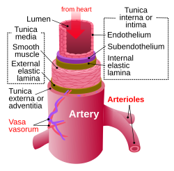
In this image, you will find the internal jugular vein, common carotid artery, subclavian vein, superior vena cava, axillary artery, pulmonary vein, brachial artery, inferior vena cava in it. Blood is pumped from the heart in the arteries. An artery is an elastic blood vessel that transports blood away from the heart. Learn the differences between an artery and a vein. Each artery is a muscular tube lined by smooth tissue and has three layers:
Anatomy Of Arteries The Diagram Of Aorta Internal Carotid Vertebrobasilar Systems And Circle Of Willis Abdominal Vascular Anatomy Abdominal Vasculature Structure Of The Aorta And Its Branches Stock Images Page Everypixel from st3.depositphotos.com Arteries are components of the cardiovascular system. Disorders or traumas affecting the common iliac arteries can have serious medical consequences. File edit view arrange extras help. The tunica medica, which is the very muscular middle layer in arteries, is thinner and less muscular in veins. A condition which arises spontaneously or as the result of trauma, where the walls of the artery are split, leading to internal bleeding and disruption of blood flow. This is the opposite function of veins, which transport blood to the heart. Heart arteries arteries and veins simple heart diagram science diagrams physical education lessons a level biology critical care nursing nursing notes circulatory system. Coronary arteries supply blood to the heart muscle.
In this image, you will find external carotid artery, internal carotid artery, vertebral artery, aorta and arch, pulmonary artery, cardiac artery, thoracic aorta, celiac trunk, superior mesenteric artery, renal artery, gonadal artery, inferior mesenteric artery, common iliac artery, external iliac artery.
John bavosi/science photo library/getty images. 5.5 / 10 ( 2 votes ) blood circulation principal veins and arteries diagram. Blood is transported in arteries, veins and capillaries. Disorders or traumas affecting the common iliac arteries can have serious medical consequences. The coronary arteries wrap around the outside of the heart. It is returned to the heart in the veins. The heart receives its own supply of blood from the coronary arteries. Arteries and veins are two of the body's main type of blood vessels. The triangles of the neck. Arteries carry blood away from the heart in two distinct pathways: File edit view arrange extras help. These arteries and their branches supply all parts of the heart muscle with blood. In this image, you will find external carotid artery, internal carotid artery, vertebral artery, aorta and arch, pulmonary artery, cardiac artery, thoracic aorta, celiac trunk, superior mesenteric artery, renal artery, gonadal artery, inferior mesenteric artery, common iliac artery, external iliac artery.
Primarily, the common iliac arteries supply blood to the bones, organs, muscles, and other structures in the abdomen and pelvis. Each artery is a muscular tube lined by smooth tissue and has three layers: 5.5 / 10 ( 2 votes ) blood circulation principal veins and arteries diagram. The main pulmonary artery splits into the right and left pulmonary arteries (better seen in the diagram at the end of this post). The narrowed arteries are at higher risk for complete blockage from a sudden.
Https Encrypted Tbn0 Gstatic Com Images Q Tbn And9gcsznadhoxopovacj8hwk Esmknahcldve Pwevncatuhzdjvsjs Usqp Cau from The capillaries connect the two types of blood. Arteries and arterioles carry oxygenated blood _____ from the heart to the body. File edit view arrange extras help. The right coronary artery courses in the right atrioventricular groove. The arteries of the head and neck. By definition, an artery is a vessel that conducts blood from the heart to the periphery. Two major coronary arteries branch off from the aorta near the point where the aorta and the left ventricle meet. Learn the differences between an artery and a vein.
28 vein artery diagram artery and vein diagram arteries and.
Primarily, the common iliac arteries supply blood to the bones, organs, muscles, and other structures in the abdomen and pelvis. Though more often occurring with carotid arteries (the other major ones supplying the brain through the neck), vertebral arteries can be impacted. Arteries carry blood away from the heart in two distinct pathways: Original vintage human anatomy victorian bookplate print 1890s medical diagram veins arteries blood circulatory system of the human body thepapermuseum. The heart receives its own supply of blood from the coronary arteries. Veins and arteries of the head (a diagram). We think this is the. Blood is transported in arteries, veins and capillaries. 13+ human body veins and arteries diagram. Arteries are components of the cardiovascular system. The two exceptions are the pulmonary and the umbilical arteries, which carry deoxygenated blood to the organs that oxygenate it (lungs and placenta. Next, we have the blood vessel responsible for carrying deoxygenated blood from the right side of the heart (right ventricle) to the lungs. You may also find the hepatic vein, portal vein, radial artery, ulnar artery.
Share :
Post a Comment
for "Arteries Diagram : Arteries Of The Spinal Cord Radiology Key"


Post a Comment for "Arteries Diagram : Arteries Of The Spinal Cord Radiology Key"